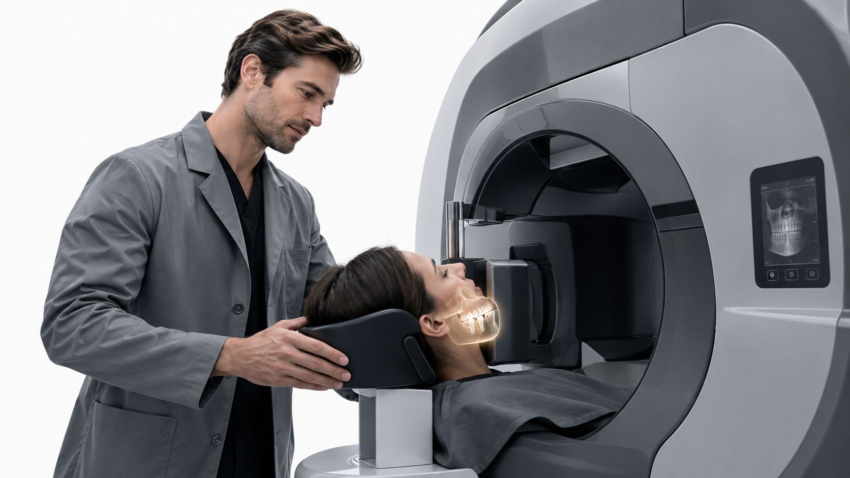
Conventional titanium dental implants are generally regarded as MRI-safe, and the FDA reports no known adverse events associated with MRI or X-ray procedures involving dental implants. The main concern is usually not the implant moving or heating dangerously, but a local image artifact that can distort nearby anatomy.
If you're in Chattanooga or Cleveland, Tennessee, and have just been told you need an MRI after getting an implant, it's understandable to wonder whether the scanner could affect your restoration or make the scan harder to read. In most cases, the practical question is what the MRI might show near the implant, not whether a fixed, integrated titanium implant makes the scan impossible.
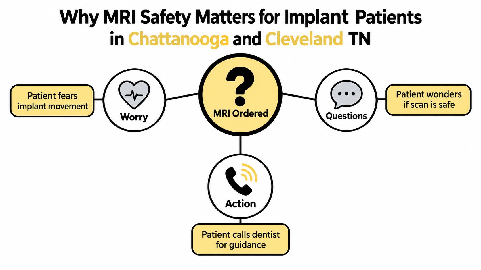
Why MRI Safety Matters for Implant Patients in Chattanooga and Cleveland TN
A head MRI can prompt a lot of questions when you have dental implants. You might picture the magnet pulling at the implant, worry that the metal will heat up, or wonder whether the scan can still show what your doctor needs to see. For conventional titanium implants, the FDA reports that it isn't aware of adverse events associated with MRI or X-ray procedures involving dental implants. It does advise patients to tell their healthcare providers and MRI technologists, because implants can distort or interfere with images (FDA guidance on dental implants and MRI).
That distinction helps put the concern in perspective. With a fixed, integrated implant, the main issue is often image clarity near the implant, particularly when the scan focuses on the jaw, face, or nearby structures. The imaging team can use your information to assess the specific components and decide how to proceed.
Safety and visibility are different questions
An MRI's powerful magnetic field and radiofrequency energy prompt legitimate screening questions. But “Will this implant make an MRI unsafe?” and “Could it affect the images?” aren't the same question. An implant might create a local shadow on an image without posing a documented safety hazard.
The type of dental restoration also matters. A permanent implant fixture isn't the same as a removable denture held in place with magnetic attachments. The MRI team needs to know what's fixed, what can be removed, and whether any parts contain magnets or an unknown alloy.
Share your implant history before imaging
Whether your MRI is ordered by a doctor in Chattanooga, Cleveland, or a nearby community, tell the imaging facility about your dental work before the appointment. If you have an implant card or records from your dentist, bring them. That information gives the MRI team a clearer picture of the materials and components involved.
The following sections distinguish fixed titanium and zirconia implants from removable magnetic overdentures, explain how material can affect image artifacts, and offer practical steps to take before your next scan.
Titanium vs Zirconia Dental Implants and MRI Artifact Size
Titanium and zirconia implants can both be compatible with MRI, but the materials don't necessarily produce the same image artifact. Artifact means a distortion or loss of detail in the image near metal or another material. It doesn't mean the implant has moved or become unsafe.
A 2020 study comparing dental implants measured a mean artifact distribution of 2.57 ± 1.09 millimeters for titanium and 0.37 ± 0.20 millimeters for zirconia. The difference was statistically significant, with p < 0.05 (2020 study comparing titanium and zirconia implant artifacts).
| Material | Mean artifact (mm) | MRI safety profile | Key consideration |
|---|---|---|---|
| Titanium | 2.57 ± 1.09 | Generally regarded as MRI-compatible, subject to screening for the specific components | May cause more local distortion than zirconia |
| Zirconia | 0.37 ± 0.20 | Generally regarded as MRI-compatible, subject to screening for the specific components | Produced a smaller measured artifact in the study |
The study also found that implant geometry didn't significantly change the artifact pattern, and the measured imaging site didn't significantly influence the result. For a patient, that means material may be more helpful than the shape of the implant post when considering potential distortion. It doesn't mean material alone can determine whether a specific MRI is appropriate.
What to do with this comparison
If you're considering dental implants near you in Chattanooga or Cleveland, ask your dentist which materials and components are planned and how they fit your clinical needs. A smaller MRI artifact can be useful when nearby anatomy may need imaging, but that's only one consideration in implant treatment. Your oral health, restoration design, and the recommendation of your dental team matter too.
And if you already have a titanium implant, a larger artifact measurement than zirconia's does not mean the implant is dangerous. It means the imaging team should know what's in place, especially when the scan targets the jaw, face, or nearby areas.
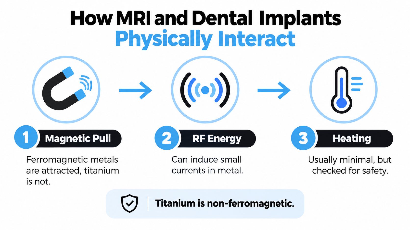
How MRI and Dental Implants Physically Interact
An MRI can interact with dental hardware in a few different ways. Understanding the difference between magnetic attraction, heating, and image distortion can make the screening process feel less mysterious.
Magnetic attraction
Conventional titanium is paramagnetic, which means it has negligible magnetic attraction compared with ferromagnetic materials. That's why a properly integrated titanium implant isn't generally expected to move because a patient enters an MRI scanner. Other dental components, particularly those with magnetic or ferromagnetic parts, need their own assessment.
Radiofrequency heating
MRI scanners use radiofrequency energy to help create images. In certain conditions, that energy can produce currents in metal and cause localized heating. Tested results offer useful context: phantom testing of titanium implants at 1.5-T and 3-T MRI reported a maximum temperature rise of approximately 0.4°C, while a separate 1.5-T study found heating below 1°C. Those levels were considered unlikely to damage nearby tissues under the tested conditions (titanium implant MRI heating results).
Under the tested conditions, titanium implant heating remained below 1°C. The MRI team still needs to check the specific implant and scanning conditions.
These results don't automatically establish that every implant system is safe under every MRI protocol. Implant design, alloy, orientation, scanner conditions, and other components can matter, so the imaging team should confirm the applicable safety information.
Image artifact
The most common practical limitation is often that the implant creates a shadow, signal loss, or distortion in nearby tissues. The closer the implant is to the anatomy being examined, the more likely it is to affect visibility. A scan focused on a distant body area may raise different image-quality concerns from one focused on the jaw or head.
The scanner's display can therefore be thought of as the place where a metal implant leaves a local mark. The implant may remain stable, while the image around it becomes more difficult to interpret.
Why Removable Magnetic Overdentures Deserve a Different Warning
A fixed implant and a removable magnetic overdenture may be described casually as “dental implants,” but they aren't the same setup for MRI screening. The implant fixture is integrated into the jaw. A removable overdenture may attach to implant-supported components with magnetic parts, which can have different MRI considerations.
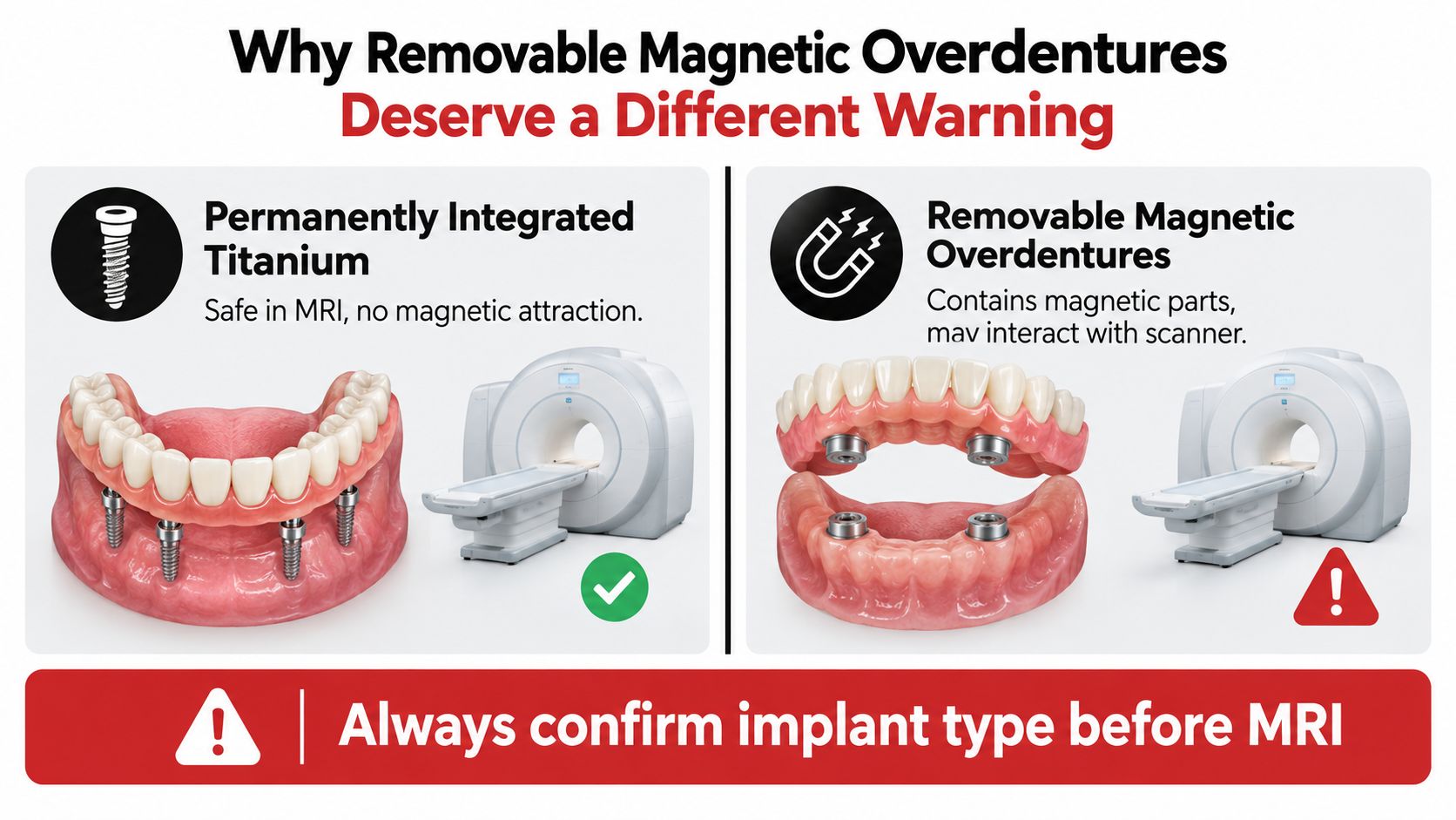
A 2025 review reported that displacement and thermal-injury risks for magnetic overdentures and implants were minimal, with measured deflection and temperature increases below critical thresholds. It also found that ferromagnetic components could produce substantial artifacts. The review concluded that fixed or hybrid titanium and zirconia prostheses are generally safe and don't need removal, while removable magnetic overdentures, especially those with an unknown alloy composition, should be removed before MRI (2025 review of MRI considerations for dental implants and magnetic overdentures).
Identify the whole restoration
The word “titanium” may describe the implant fixture, but it doesn't necessarily tell you what materials are used in the abutment, crown, bar, or removable attachment. If you wear an implant-supported denture, be clear about which parts come out and whether it uses magnetic retention.
For patients learning about denture implants in Chattanooga, understanding how a restoration attaches can also help with conversations about future imaging. The MRI facility should make the final decision about whether an appliance or component can remain in place during a scan.
Practical rule: Don't assume every part of a dental restoration has the same MRI requirements. Tell the imaging team about fixed implants, removable prostheses, and any magnetic attachments separately.
The review noted limitations in the available evidence, including small sample sizes and varied testing methods. That's another reason not to rely on a blanket statement such as “titanium means everything is safe.” Describe what you have, bring records if available, and follow the MRI facility's instructions.
Your Pre-MRI Checklist for Dental Implant Patients
A little preparation can help the MRI team assess your dental work without guesswork. If you're unsure what kind of restoration you have, your dentist may be able to help identify it, but the MRI facility determines whether a particular device is acceptable under its scanning conditions.
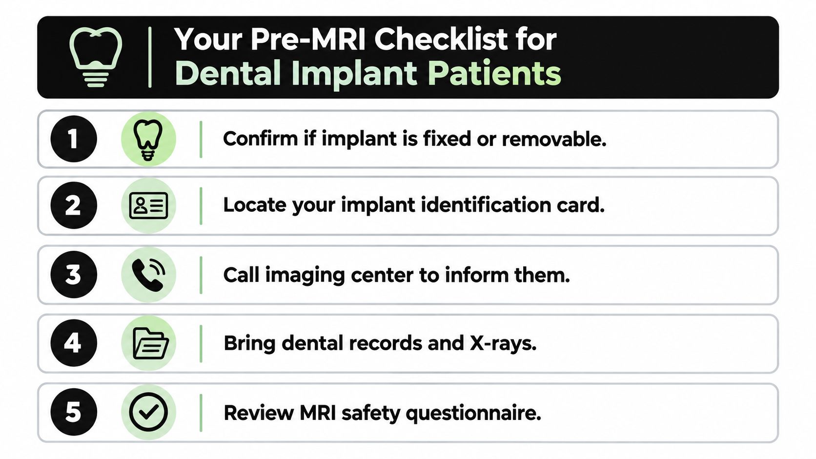
Confirm what's fixed and what's removable. Identify whether you have a fixed implant restoration, a removable overdenture, or both. Tell the imaging team if the removable part uses magnetic attachments.
Look for implant records. Bring an implant card, treatment records, or other documentation that identifies the components and materials. A review of metallic dental prostheses describes titanium's paramagnetic behavior as supporting its MRI safety, while noting that image artifacts, heating, and other materials can still matter (review of metallic dental prostheses and MRI).
Call the imaging center before your appointment. Tell the staff about your implant fixture, abutment, crown, removable prosthesis, magnetic attachments, or orthodontic hardware. The FDA advises patients to notify healthcare providers and MRI technologists because dental implants may distort or interfere with images.
Ask about image quality. If the scan is focused on the head, jaw, or face, ask whether the radiology team can use metal-artifact-reduction sequences or whether another type of imaging may better answer the clinical question.
Follow the facility's instructions. Removable appliances may need to come out, but don't remove or alter a fixed restoration yourself. The MRI team will review your screening information and make the scan-specific decision.
If you're weighing imaging options for a separate concern, this plain-language guide to MRI or X-ray for your back can help you understand how clinicians may use different scans. For questions about implant treatment and documentation, a dental implant treatment plan can also help you keep track of the components involved.
How a Brain or Head MRI Can Still Work With Dental Implants
A dental implant doesn't automatically prevent a head or brain MRI. If the scan is looking at the brain, the radiology team can consider where the implant sits and whether any artifact is likely to interfere with the area they need to assess.
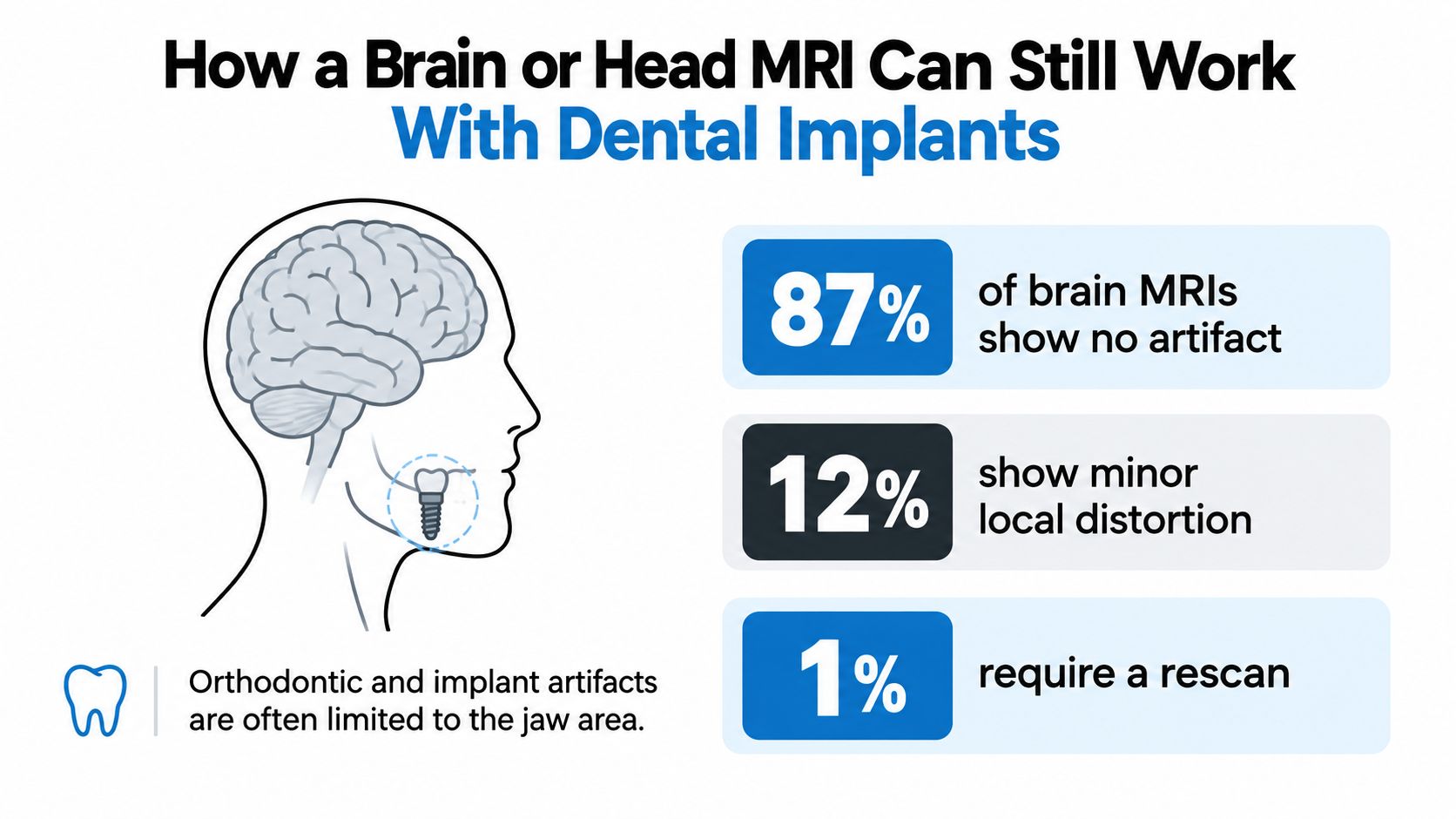
A study of brain MRI artifacts reported that orthodontic appliances accounted for 78% of observed artifacts, titanium dental implants for 18%, and metallic crowns for 4% (study of dental artifacts in brain MRI). These figures describe the distribution of observed artifacts in that study. They don't mean that implants prevent a corresponding share of brain scans or that every patient will have the same image result.
Help the radiology team plan
Artifact depends on the implant material and location, as well as the MRI settings and the anatomy being examined. If the scan targets the brain, a local distortion near the jaw may not affect the part of the image the clinician needs. The radiologist can assess whether the images remain useful and whether an adjustment could help.
Before the appointment, tell the imaging staff where your implant is and provide the implant records if you have them. If you're a patient in Cleveland or Chattanooga, your dental office may be able to document the implant location and available component details for your care team. That information can help the radiology staff plan the scan, though the imaging facility still makes the final decision.
A practical example: a patient with a fixed implant who is scheduled for a brain MRI can alert the imaging center in advance, bring the implant card, and answer questions about any removable dental appliance. The technologist can then review the screening details rather than discovering the restoration midway through the appointment.
Talking With Winn Smiles Before Your Next MRI Referral
Your dentist can help you understand what was placed in your mouth and provide records, but the MRI facility must decide whether the specific implant and scanner conditions are compatible. A clear handoff helps both teams work from the same information. The FDA advises notifying healthcare providers about dental implants because they can interfere with images, and reports no known adverse events associated with MRI or X-ray procedures involving dental implants (review discussing FDA guidance on dental implants and imaging).
Ask for details you can share
If you're a Winn Smiles patient in Chattanooga or Cleveland, contact the practice before an imaging appointment if you need help identifying your dental components. Ask whether your records describe the implant fixture, abutment, crown, and any removable prosthesis or magnetic attachment. Keep the information with your personal health records and share it with the MRI technologist.
That conversation can be especially helpful if you don't know whether a denture is magnet-retained or can't remember which parts are removable. Don't rely on the word “titanium” alone to describe an entire restoration. Give the MRI facility all the information you have, including uncertainty about a material or attachment.
Dental implant care is part of your broader oral health, alongside cleaning and exams, restorative dentistry, and emergency dental services when needed. If you're looking for a dentist in Chattanooga or Cleveland, or considering dental implants near you, you can ask about documentation during a consultation. A local dental team can also help you understand the restoration and how to describe it to other healthcare providers.
For an MRI, the essential approach is straightforward: identify what's fixed and removable, gather your records, and tell the imaging staff before the scan. If you need help reviewing your implant details or discussing treatment options, schedule a visit with Winn Smiles in Chattanooga or Cleveland.
Winn Smiles provides dental implant consultations, full-arch implant care, restorative dentistry, and help understanding your implant records before an MRI referral. Contact Winn Smiles to schedule a consultation or request a second opinion in Cleveland or Chattanooga.